The Undergraduate Instrumentation Facility supports the department by providing essential scientific instruments, equipment, and dedicated staff. Its mission is to maintain research-grade instruments, offer comprehensive training, and promote a safe working environment. The facility prioritizes the acquisition of cutting-edge technology, the integration of advanced tools into teaching laboratories, and the delivery of hands-on experience for students. Additionally, it supports both faculty and student research, helping students develop valuable research skills throughout their academic journey.
The Instrumentation Facility takes a proactive approach to the maintenance and acquisition of advanced scientific instruments, aligning with current technological trends and advancements. Our goal is to integrate high-tech instrumentation into our teaching laboratories to offer students direct, hands-on experience in laboratory courses; to provide training that ensures the safe and effective use of the equipment; and to share our expertise in support of student and faculty research. This approach allows students to work with cutting-edge instruments and gain valuable research experience as they progress through our academic programs.
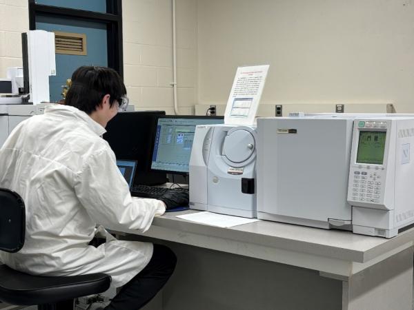
GC-MS is a powerful analytical technique used to identify and quantify chemical compounds at low detection limits in a wide range of samples, including food, pharmaceuticals, fuels, and environmental sources. In gas chromatography (GC), volatile compounds in a mixture are separated by passing them through a column carried by an inert gas. Each compound elutes at a different time depending on its interaction with the column material. The mass spectrometer (MS) then detects these compounds by ionizing them and generating ion fragments. The resulting mass and fragmentation patterns are used to identify the compounds. GC-MS combines these two analytical tools and is widely employed in pharmaceutical development, forensic science, and various research applications.
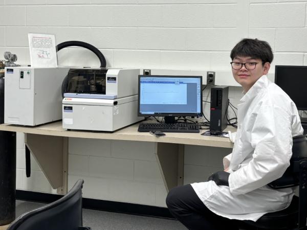
DSC measures the amount of heat required to maintain the temperature of the sample the same as that of an inert reference material as both are heated or cooled at a constant rate. During phase transitions, such as, melting, decomposition, or structural changes, the sample will either absorb or release heat, requiring more or less energy than the reference to maintain the same temperature. This allows DSC to precisely determine both the temperature at which these transitions occur, and the amount of energy involved in the process. DSC is commonly used in polymer chemistry, materials science and the analysis of material purity.
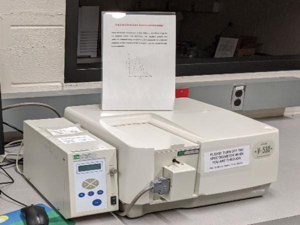
The instrument measures the energy required to excite the electrons in molecules by analyzing the amount of light absorbed at different energy levels. This technique, commonly used in compound analysis, is based on the principle that the amount of light absorbed is directly proportional to the concentration of a substance in solution. It is widely used to characterize, identify and quantify compounds across various fields, including chemistry, biology, and environmental science.
The instrument functions similarly to a conventional UltraViolet-Visible (UV-Vis, 200-400nm) spectrometer but offers an extended wavelength range (200-2500 nm). It is designed to capture spectra from both liquid and solid samples. This enhanced versatility makes it particularly valuable for analyzing materials that are challenging to dissolve, thereby expanding its potential applications across various fields.
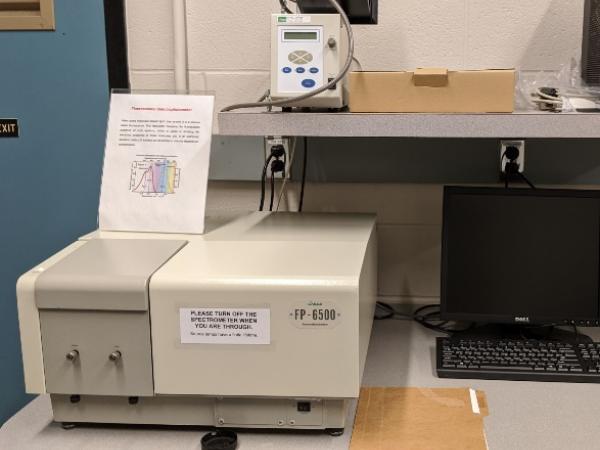
This instrument measures the light re-emitted by some molecules when they absorb light – a process known as fluorescence. It captures the fluorescence spectrum of these systems, providing valuable insight into the electronic properties of these molecules. Because emission intensity is directly related to concentration, fluorescence spectroscopy offers exceptional sensitivity, making it a powerful tool for precise quantitative and qualitative analysis
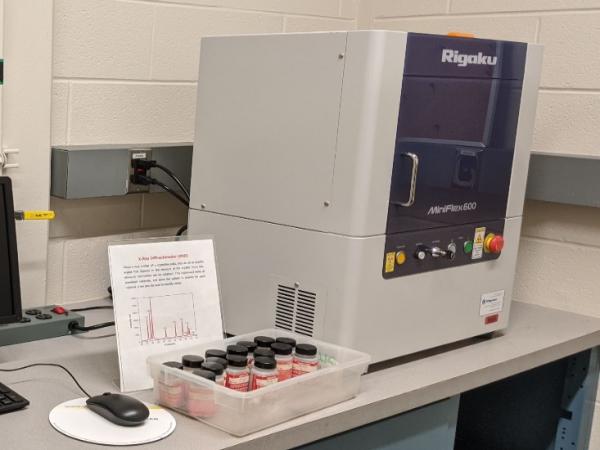
X-ray diffraction (XRD) is an analytical technique used to investigate the structure of materials by measuring how X-rays scatter when they interact with a crystalline solid. The resulting diffraction pattern provides information about the atomic arrangement within the crystal, allowing for the determination of structural features such as atomic spacing, crystallite size, and symmetry. XRD is widely used in material science, chemistry, and geology to identify and characterize crystalline substances.
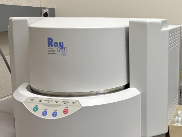
An XRF spectrometer is an X-ray instrument used for non-destructive chemical analysis of materials such as rocks, minerals, sediments and fluids. It operates by exciting the inner electrons of atoms, prompting the emission of fluorescent X-rays with energies characteristic of each element. The instrument measures these fluorescent X-rays to identity the elements in a sample, and sometimes determine their quantitative composition. Unique to this method is its nondestructive nature allowing valuable samples such as jewelry, artworks, and archaeological artifacts to be analyzed without damage.

An NMR instrument is used to analyze the molecular structure of materials by measuring the interaction of nuclear spins in a powerful magnetic field. In nuclear magnetic resonance (NMR), certain nuclei exist in specific spin states when exposed to an external magnetic field. NMR detects transitions between these spin states, which are unique to the nuclei and their chemical environment, allowing for compound identification. NMR is widely used in industries like medicine, biochemistry, and physics.
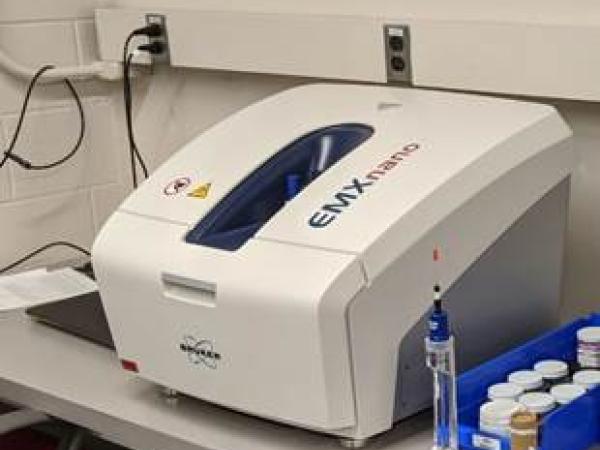
EPR is a technique used to study chemical species with unpaired electrons, such as organic and inorganic radicals, transition metal complexes, and some biomolecules. Electrons usually prefer to pair, but unpaired electrons exist in systems like free radicals. EPR can excite these unpaired electrons and generate spectra that reveals information about the atom hosting the unpaired electrons, its surrounding geometry, and neighboring atoms, making it useful for structure determination. EPR is also commonly used to estimate the shelf life of products containing free radicals, such as tea, coffee, bagels, and beer.
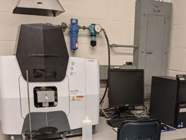
This instrument is used for trace analysis, typically in parts-per-million range. The sample is introduced into the instrument and passed through a high-temperature flame that breaks down compounds into individual atoms. Each element absorbs light at specific wavelengths, and by measuring the absorption, the concentration of the elements in the solution can be determined. The degree of absorption is directly related to concentration, enabling accurate quantification of trace elements.
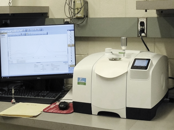
FTIR is an analytical instrument that measures the energies associated with the vibrations of atoms within a molecule, specifically in the infrared region of the electromagnetic spectrum. These vibrational energies are characteristic of the bonds and structural features of the molecule. By analyzing these vibrational frequencies, FTIR allows for the determination of the molecular structure, making it a powerful tool for molecular identification and characterization.
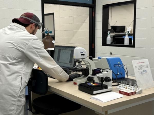
A Raman spectrophotometer analyzes how visible light interacts with a molecule, either being absorbed or scattered. In scattering, some photons transfer energy to the molecule, exciting its vibrational modes, which are recorded in the Raman spectrum. Unlike infrared spectra, Raman spectra are based on scattering, offering complementary vibrational information not seen in infrared spectra. Both techniques can be used together for structural determination and compound identification. Raman spectroscopy is non-destructive and is especially useful for pigment identification in artwork, aiding in conservation and study of historical pieces.
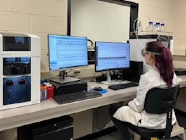
This is a technique that separates charged molecules, such as proteins, within a narrow capillary tube using an electric field. The separation occurs in an aqueous buffer, where the sample migrates at different rates depending on its size, charge, and electrochemical properties. CE is highly advantageous for protein analysis due to its high resolution, speed, and sensitivity. It is ideal for analyzing complex biological samples, providing insights into protein heterogeneity, molecular weight, and charge variations, while requiring small sample volumes and minimizing sample degradation.
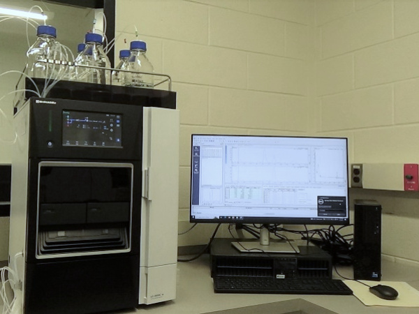
Liquid Chromatography (LC) separates and analyzes components in a liquid mixture by passing the sample through a column with a stationary phase. Components separate based on interactions like polarity, size, or charge. High-Performance Liquid Chromatography (HPLC) is an advanced LC variant using higher pressures for faster, more precise separations, commonly used in chemical, pharmaceutical, environmental, and food analysis.
